Impacted Canine Treatment in Austin

Impacted canines treated at Limestone Hills follow a predictable surgical-orthodontic sequence: 3D CBCT planning to locate the canine in three dimensions, periodontist or oral-surgeon exposure with a chain or attachment bonded directly to the tooth, then 6 to 18 months of orthodontic traction to bring the canine into the arch. The 3D scan at the free consultation locates the canine and identifies which neighboring roots are at risk so the eruption pathway is planned before any surgical work.

  • PhD Orthodontist
  • 4.9★ 220+ Reviews
  • Mon-Sun 7:30 AM - 10:30 PM
  • Oral Surgery Coordination
*For new patients only. Patients in treatment $150, deductible from comprehensive treatment fee.
Treatment plan for impacted canine at Limestone Hills Orthodontics in Austin, TX, with braces, chain, and tooth movement.
Home / Problems / Impacted Canine

What Is an Impacted Canine?

An impacted canine is a permanent canine tooth that has failed to erupt into its correct position in the dental arch by the expected age (typically age 12 to 14 in girls and 13 to 15 in boys).

The most common impaction site is the upper canines (about 2 percent of the population), with the canine displaced palatally (toward the roof of the mouth, in roughly two-thirds of cases) or buccally (toward the cheek, in the remaining third). Lower-canine impaction is much rarer.

The clinical concern is not aesthetic. An impacted canine that is left in place tends to drift over years, resorbing the roots of the adjacent lateral incisor or first premolar. Permanent root loss in those neighbors is the worst-case outcome, and it is silent and progressive without 3D imaging.

Limestone Hills screens for canine impaction at every Phase 1 evaluation around age 9 to 11 specifically because catching the problem before the canine is fully impacted dramatically simplifies treatment and reduces the risk of root resorption damage.

Why Canine Teeth Are Worth Saving

Modern orthodontics nearly always favors saving the impacted canine over removing it for two structural reasons. The canine is the longest-rooted tooth in the mouth, with a deep bony attachment that anchors the corner of the arch and supports the lip and cheek architecture.

Replacing it cosmetically with a premolar substitution or a dental implant produces an arch that is mechanically weaker and aesthetically asymmetric in roughly half of cases (the canine eminence visible at the corner of the smile cannot be reproduced by either substitute).

The canine also leads occlusion in lateral movements (canine guidance), distributing chewing forces across the arch in a way that protects the molars from cusp-tip wear. Premolar substitution shifts that load onto teeth the dentition was not designed to bear in lateral function.

Patients in their 50s and 60s whose canines were extracted in adolescence often present with asymmetric posterior wear and TMJ symptoms that trace back to the missing canine guidance.

The Limestone Hills Approach to Impacted Canines

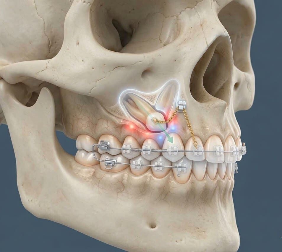
Three-dimensional imaging first. An impacted canine cannot be treated correctly without CBCT. On a 2D panoramic x-ray, a canine trapped in the palate looks nearly identical to a canine trapped in the buccal (cheek) side, and the treatment is different. Once position is clear, Dr. Viecilli works with the oral surgeon on exposure and bonds a chain to the tooth, then pulls it into the arch along a biomechanically sensible path. Recovery, root length, and adjacent-tooth damage depend on the approach chosen before the surgery starts, which is why the imaging and planning step matters more than the surgical step.

The practical sequence runs about 9 to 18 months depending on canine position.

CBCT planning at the consultation, surgical exposure with a periodontist or oral surgeon (typically a 30-minute outpatient procedure under local anesthesia), and orthodontic traction during comprehensive braces or aligner treatment that brings the canine through the bone and into its correct position.

TADs (temporary anchorage devices) are used routinely as the traction anchor so the rest of the dentition is not dragged toward the impaction site during the long pull-down phase.

Expose-and-Bond vs. Premolar Substitution

The decision between expose-and-bring-down (preserve the canine) and extract-and-substitute (replace with a premolar or implant) is made on three findings from the CBCT scan:

Position and angulation. A canine angled less than 45 degrees from vertical and within 5 millimeters of the occlusal plane has a strong prognosis for orthodontic traction. A canine angled more than 60 degrees or displaced more than 8 millimeters from the arch has a markedly worse prognosis and may not be worth saving when the patient is post-growth.
Damage to neighboring roots. Significant resorption already present on the lateral incisor or first premolar may force a different plan: either accept the resorption damage and bring the canine down anyway, extract the resorbed neighbor and use the canine in its place, or extract the canine and restore.
Patient age and ankylosis risk. Adolescent and young-adult cases tolerate traction well; patients over age 30 with long-standing impaction face higher ankylosis risk and may need a different approach.
Premolar substitution remains the right answer in a minority of adult cases where the canine prognosis is genuinely poor or the lateral incisor has been lost. Dr. Viecilli discusses both pathways openly at the consultation and shows the CBCT imaging on-screen so the patient understands the structural tradeoff before committing to either route.

Early Detection: The Age 9–10 Window

The single most consequential intervention in canine-impaction management is early detection. By age 9 or 10, panoramic and CBCT imaging show the path the canine is tracking through the alveolus toward the arch. Three interventions during the active growth window (ages 9 to 12) can each redirect a canine that is heading toward impaction:

Extraction of the deciduous (baby) canine at the right moment can give the permanent canine the eruption pathway it needs. Several published studies show this single intervention resolves about half of developing palatal impactions when performed at age 10 to 11.
Palatal expansion in cases where the upper arch is narrow can create the transverse space the canine needs to navigate the arch correctly. Expansion during the open-suture window (before age 14) is the highest-leverage version of this intervention.
Distal-driving mechanics with a limited orthodontic appliance can move the first molar back to recover lost arch length when the primary teeth have been lost early or the permanent first molars have drifted mesially.

Limestone Hills evaluates every Phase 1 patient for canine eruption pathway specifically because the cost-benefit math favors early intervention by a wide margin: a 3-month deciduous-canine extraction at age 10 is incomparably simpler than a 12 to 18 month surgical-exposure-and-traction sequence at age 14.

What Happens If an Impacted Canine Is Not Treated

Left untreated, an impacted canine carries three structural risks that compound over time:

Root resorption of the adjacent lateral incisor (the tooth next to where the canine should be). Studies show 30 to 70 percent of palatally impacted canines produce some degree of root resorption in the lateral incisor, and the damage is silent and progressive until the lateral incisor becomes mobile or is lost. The 3D CBCT scan at consultation is the only imaging that reliably visualizes early resorption.
Cyst formation around the impacted tooth follicle. Dentigerous cysts form in roughly 1 to 2 percent of long-impacted canines and require surgical removal once detected. Catching the impaction before cyst formation avoids the surgical morbidity entirely.
Ankylosis (fusion of the canine to the surrounding bone). The longer a canine remains impacted, the higher the risk of ankylosis, which permanently prevents orthodontic traction and forces extraction. Ankylosis risk rises significantly in patients over age 25, which is why adult-presenting long-impacted canines often have a worse prognosis than adolescent cases.

The Impacted Canine Treatment Process at Limestone Hills

1

CBCT Localization

3D imaging determines the exact position, angulation, depth, and relationship to adjacent roots. This guides the surgical approach and the direction of orthodontic traction.

2

Create Space

Braces are placed and the teeth are aligned to create a gap in the arch where the canine belongs. If the arch is narrow, palatal expansion may be needed first. The baby canine is extracted if still present.

3

Surgical Exposure

An oral surgeon lifts the gum tissue, removes any overlying bone, and bonds an orthodontic bracket with a small chain onto the impacted canine. Closed technique (gum replaced) or open technique (gum left open) is chosen based on the tooth’s position. Takes approximately 1 hour under local anesthesia.

4

Orthodontic Traction

Dr. Viecilli uses the chain to apply light, slow traction. Gradually guiding the canine through the bone and into its correct position over 6–12 months. Force must be light to avoid ankylosis (tooth fusing to bone) or root resorption.

5

Final Alignment

Once the canine has erupted into the arch, comprehensive braces fine-tune its position, establish proper canine guidance, and align the rest of the teeth. Total treatment: 18–30 months.

Why Limestone Hills Handles Impacted Canines Carefully

Three points distinguish the Limestone Hills approach to impacted canines from a generic surgical-orthodontic referral:

3D CBCT planning before the surgical appointment, not after. The exposure surgeon needs to know the canine’s exact position, angulation, and proximity to neighboring roots BEFORE making the incision. Limestone Hills includes the CBCT in the consultation and shares the imaging directly with the surgical partner so the surgical plan is built on three-dimensional data rather than a panoramic estimate.
TAD anchorage during traction by Dr. Viecilli in-office. Conventional traction anchors against the rest of the dentition, which dragsthe arch toward the impaction site over months. A TAD anchored in palatal or alveolar bone holds the rest of the dentition stable while the canine is pulled into place. No second surgical office visit, no separate TAD-placement fee for comprehensive cases.
Early-detection screening at every Phase 1 consultation. The cheapest impacted-canine case is the one that never becomes impacted because the deciduous canine was extracted at age 10. Limestone Hills screens for canine eruption pathway on every panoramic and CBCT scan and recommends the simple early intervention openly when the imaging supports it.

Treatment Timeline

Stage Duration What Happens
Screening & imaging 1–2 visits Panorex, CBCT 3D localization
Interceptive (ages 10–11)
6–12 months
Baby canine extraction, monitor eruption
Space creation
4–8 months
Braces, expansion if indicated
Surgical exposure
1 day procedure
Oral surgeon exposes and bonds bracket
Traction + alignment
12–18 months
Light traction to guide canine into arch
Detailing & retention
3–6 months
Establish canine guidance, retainers

What to Expect at Your Consultation
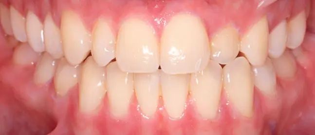
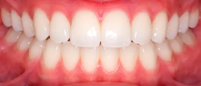
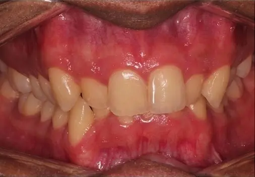
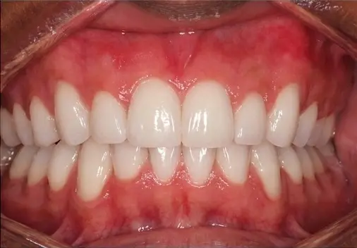
Digital intake. Dental history, presence of baby canines, any noticed asymmetry.
Comprehensive records. Digital scan, clinical photos, panoramic x-ray, free CBCT for 3D localization.
Diagnosis with Dr. Viecilli. Canine position, angulation, impact on adjacent teeth.Diagnosis with Dr. Viecilli. Canine position, angulation, impact on adjacent teeth.
Decision: save vs. substitute. Honest discussion of both paths with risks and timelines.
Exact pricing. Including oral surgeon coordination and insurance pre-authorization.

Before & After

Showing clean, straight teeth and healthy gums after crossbite correction at Limestone Hills Orthodontics in Austin, TX. At Limestone Hills Orthodontics in Austin, TX, a close-up of a mouth shows clean, white teeth and healthy gums after treatment.
Before
After

Crowding: Invisalign/Clear Aligners

Crowded, misaligned teeth with visible gums shown before treatment at Limestone Hills Orthodontics in Austin, TX. A patient at Limestone Hills Orthodontics in Austin, TX shows straight, white teeth and healthy gums after overbite correction.
Before
After

Deep Overbite: Braces with Bite-Pads

See more impacted canine cases in our treated cases gallery.

How Much Does Impacted Canine Treatment Cost?

Comprehensive braces (includes canine management): from $4,000
Clear ceramic braces: from $4,600
Surgical exposure: separate oral surgeon fee, typically $800–$1,500 per tooth

The practice coordinates directly with the surgeon for scheduling and insurance pre-authorization. Most dental insurance covers both the orthodontic treatment and the surgical exposure when medical necessity is documented (impacted tooth, risk of root resorption).

Insurance & Financing

OrthoSync: 0% interest in-house financing, no credit check.
Cherry: $189.99 down with a soft credit pull.
CareCredit: $0 down with approved credit.
Pay-in-Full: 3% discount applied automatically.

Most PPO orthodontic plans cover the orthodontic component (traction, comprehensive treatment); the exposure surgical fee is billed by the partnering periodontist or oral surgeon and is often covered separately under medical insurance. The free CBCT scan at consultation is included.

Common Appliances Used at Limestone Hills

Guiding an impacted canine into position requires fixed braces to hold space open and direct the tooth’s path of eruption over 12 to 18 months; when the anchorage demands of the case are high, temporary anchorage devices give Dr. Viecilli precise control without relying on patient cooperation.

Frequently Asked Questions About Impacted Canines

How do I know if my child has an impacted canine?

Check for asymmetric canine bulge. Feel the palate near the gumline on both sides. If one side has a bump and the other doesn’t, imaging is warranted. Also: baby canine still present after age 13, visible gap, or asymmetry between left and right. Panoramic x-ray at age 9–10 can identify ectopic canines early.

What happens if an impacted canine is not treated?

Root resorption of adjacent teeth (lateral incisor most at risk. Silent, no symptoms until damage is severe), dentigerous cyst formation, eventual loss of the baby canine leaving a gap, and progressive arch collapse.

Can an impacted canine erupt on its own?

If caught early, extracting the baby canine removes the obstruction and allows self-correction in 62–78% of palatal cases. Once fully impacted, surgical exposure and orthodontic traction are needed.

What is the expose and bond procedure?

Oral surgeon lifts gum tissue, removes overlying bone, bonds a bracket + chain onto the impacted canine. Approximately 1 hour under local anesthesia. Orthodontist then uses the chain to guide the tooth into position over 6–12 months.

Is the surgery painful?

Done under local anesthesia with sedation available. Post-op discomfort is mild to moderate. Generally less than wisdom tooth extraction. OTC pain medication for 2–3 days. Soft diet for one week.

How long does the entire treatment take?

18–30 months depending on canine position. Space creation: 4–8 months. Surgical exposure: 1 day. Traction and alignment: 12–18 months.

When is extraction the better option?

When the canine is in an extreme position, is ankylosed (fused to bone), or in older patients where guided eruption has a poor prognosis. The first premolar is substituted in the canine position and reshaped.

Related Conditions

Crowding

Gaps & Spacing

Crossbite

Children’s Orthodontics

Overbite